In-Gel Westerns
In-Gel Westerns directly detect protein in the polyacrylamide gel, without membrane transfer or blocking. Near-infrared (NIR) fluorescent In-Gel Westerns can be imaged with the Odyssey® M or Odyssey DLx Imagers when using IRDye® secondary antibodies for detection.

In-Gel Westerns are useful for:
- Large, hydrophobic, or post-translationally-modified proteins, such as glycoproteins, receptors, or cell membrane proteins that do not transfer well
- Small proteins that may pass through the membrane during transfer
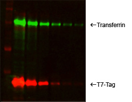
- Multiplexed, two-color detection of two different protein targets within the same gel
Advantages of Near-Infrared In-Gel Westerns
- Save time and reduce cost of running a blot
- Eliminate variables associated with membrane transfer and blocking
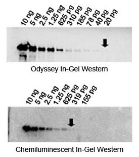
- NIR In-Gel Westerns provide sensitivity comparable to chemiluminescent In-Gel detection with clearer, sharper bands (Figure 2), but not as high as traditional NIR membrane Western detection.
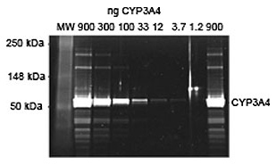
- Detect to nanogram to picogram levels (Figure 3)
- Produce clean images with minimal background banding


Typical In-Gel Western Workflow
Perform electrophoresis

Remove stacking gel

Add fixation solution to fix proteins in gel

Wash

Incubate with primary antibody

Wash

Incubate with secondary antibody

Wash

Excite with infrared lasers

Acquire image using Odyssey Imager and analyze

References
- Theisen, M. J. and Chiu, M. L. In-gel immunochemical detection of proteins that transfer poorly to membranes.
