BrightSite™ IRDye® in vivo Imaging Agents
Products
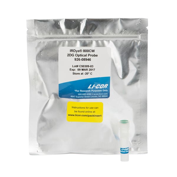
IRDye® 800CW 2-DG Optical Probe
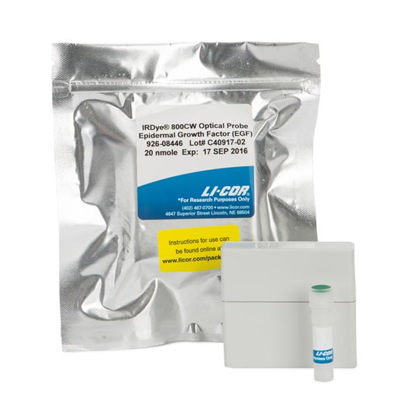
IRDye® 800CW EGF Optical Probe
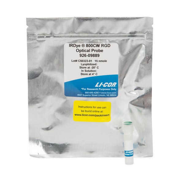
IRDye® 800CW RGD Optical Probe
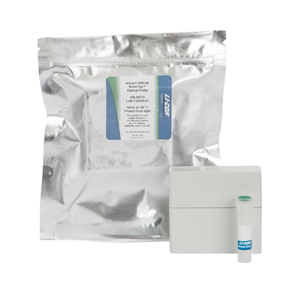
IRDye® 800CW BoneTag™ Optical Probe
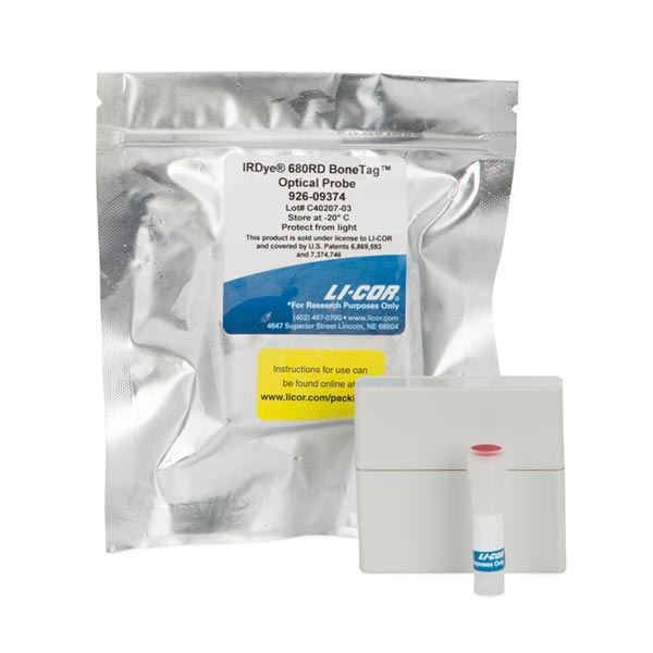
IRDye® 680RD BoneTag™ Optical Probe
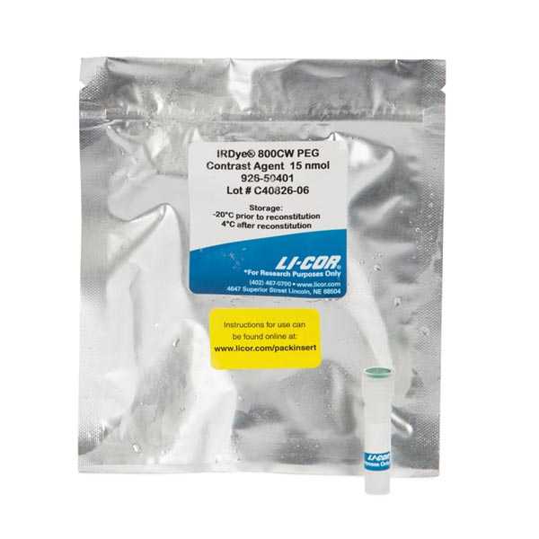
IRDye® 800CW PEG Fluorescent Contrast Agent
Every LI-COR BrightSite IRDye in vivo imaging agent has been carefully validated with cultured cell assays, microscopy, in vivo imaging of animal models, and histology to ensure high affinity and specificity.
BrightSite IRDye Small Animal Imaging Agents are:
- Ready-to-use probes that are simple to administer, allowing you to begin animal studies immediately
- Sensitive, offering the high signal-to-noise advantages of near-infrared fluorescence which are especially important for small or deep targets
- Cited in several hundred peer-reviewed publications so you can use them with confidence
- Imaged with any small animal imaging equipment with appropriate 680 nm or 800 nm filter sets
- Compatible with most small animal imaging systems, including instruments from LI-COR Biosciences (Pearl® and Odyssey®), PerkinElmer (Xenogen, Caliper, CRi, and VisEn), and Bruker (Carestream and Kodak)
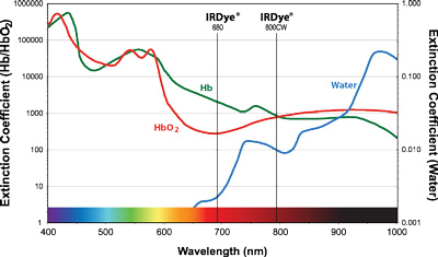
IRDye 800CW absorption/emission near 800 nm matches near-infrared absorption minima for bodily fluids and tissues, resulting in excellent tissue penetration, making it ideal for in vivo imaging.
Fluorescently Label Cells or Tissues for in vivo or in vitro Studies
CellVue® fluorescent imaging kits use proprietary labeling technology to stably incorporate fluorescent dyes containing long aliphatic hydrocarbon tails into lipid membranes.1 They are useful for researchers working in all aspects of science and technology where fluorescently-labeled cells and/or tissues are required. CellVue dyes also provide researchers with valuable tools for many in vivo and in vitro cell studies using fluorescent membrane labels.
CellVue dyes consist of long aliphatic hydrocarbon tails linked to a polar fluorescent chromophore.
- These extremely lipophilic fluorescent dyes rapidly and stably integrate into the phospholipid membrane of cells or other membrane-containing bioparticles by non-covalent interactions
- The dyes are stably maintained within the lipid bilayer through strong hydrophobic interactions and do not transfer into the unstained membranes of adjacent cells, which permits a labeled cell to be tracked for extended periods of time
- The rapid incorporation of the dye allows for immediate analysis of cell functions without a waiting period
Selecting the Right Optical Imaging Agent
| Application | IRDye EGF | IRDye RGD | IRDye 2-DG | IRDye PEG | IRDye BoneTag | CellVue |
|---|---|---|---|---|---|---|
| Tumor Imaging | ||||||
| Metabolic Imaging | ||||||
| Inflammation/Arthritis | ||||||
| Vasculature (Contrast) | ||||||
| Lymphatic Imaging | ||||||
| Lymph Node Imaging | ||||||
| Structural Imaging | ||||||
| Cell Trafficking |
References
- Horan, P. K., and Slezak, S. E. (1989) Nature, 340. 167-168.