Primary Antibodies for Normalization
Products
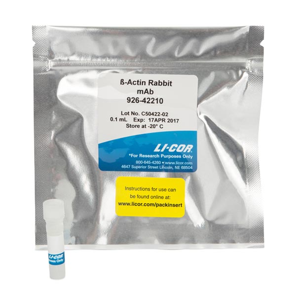
β-Actin Rabbit Monoclonal Antibody
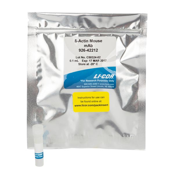
β-Actin Mouse Monoclonal Antibody
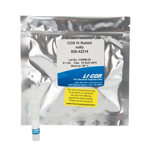
COX IV Rabbit Primary Antibody
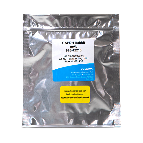
GAPDH Rabbit Monoclonal Antibody
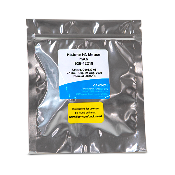
Histone H3 Mouse Monoclonal Antibody
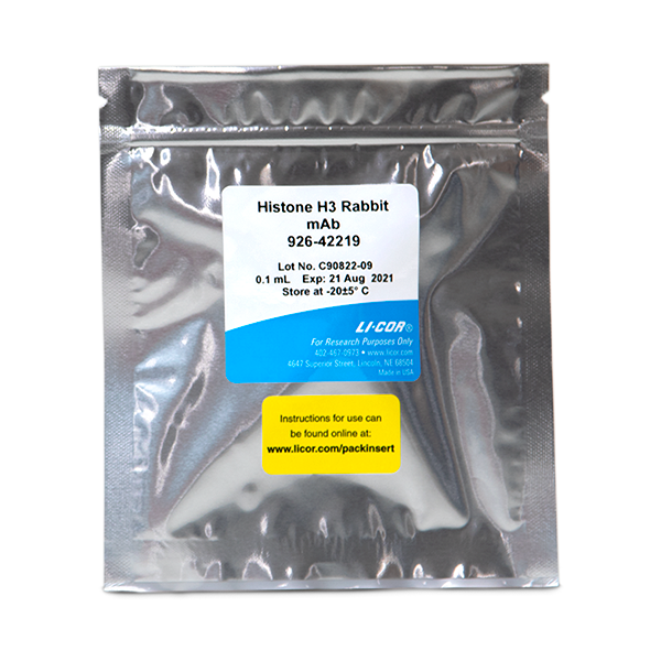
Histone H3 Rabbit Monoclonal Antibody
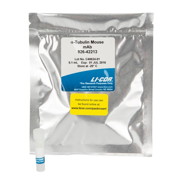
α-Tubulin Mouse Monoclonal Antibody
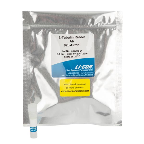
β-Tubulin Rabbit Polyclonal Antibody
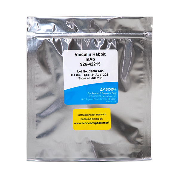
Vinculin Rabbit Monoclonal Antibody
When performing quantitative Western blots, internal loading controls and normalization are essential for reliable, precise comparison of protein expression. LI-COR offers several primary antibodies which can be used for two-color normalization when performing multiplex Western blots or for cell-based assay normalization, such as for In-Cell Western™ Assays.
For Western blots, lysate preparation, sample loading, and membrane transfer introduce unavoidable variation. After validation, housekeeping proteins (HKP) can be used for normalization of protein levels.
- Normalize and correct for uneven loading, using the intensity of the internal loading control
- Visually compare protein levels with confidence, even if you don't quantify bands
- Ensure that visually-observed changes in protein levels represent actual change, not artifacts
Read more about using a housekeeping protein as an internal loading control.
When using an HKP as your normalization strategy, it’s important to validate your HKP for each experiment to ensure its expression is stable. Many factors can influence expression including tissue, treatment, and cell density.
"'House-keeping' proteins should not be used for normalization without evidence that experimental manipulations do not affect their expression."
The Housekeeping Protein Validation Protocol can help you validate that your HKP expression is not changing with treatment. The Housekeeping Protein Normalization Protocol will guide you through the right experimental steps and calculations for normalization using an HKP. Validating your HKP is the only way to know that expression is not affected by the conditions of your experiment
For In-Cell Western Assays, a second protein target (such as actin, tubulin, COX IV, or GAPDH) can be used for normalization. Abundance of the normalization target must be unaffected by the cell treatments used.
Learn more about ICW assay normalization methods.
Comparison of Housekeeping Loading Controls
The table below has a few examples of some common housekeeping proteins, their molecular weights, and a few considerations. This list is not complete, and any housekeeping protein you choose should be fully validated for your system under study.
| Housekeeping Protein | Molecular Weight | Note |
|---|---|---|
| COX IV | 17 kDa | Excellent choice for low-abundance target proteins. |
| Histone H3 | 17 kDa | Not suitable for experiments where the nuclear envelope has been removed. |
| PCNA | 36 kDa | PCNA is quickly degraded when DNA damage pathways are activated.1 |
| GAPDH | 36-38 kDa | |
| β-Actin | 45 kDa | |
| β-Tubulin | 52-55 kDa | Not suitable for comparing leukocytes from different age groups.7 |
| Lamin B1 | 68 kDa | Not suitable for experiments where the nuclear envelope has been removed.7 |
| Vinculin | 124 kDa | Can be used as a loading control for high molecular weight proteins.7 |


References
- Cazzalini O, Sommatis S, Tillhon M, Dutto I, Bachi A, Rapp A, Nardo T, Scovassi AI, Necchi D, Cardoso MC, Stivala LA, Prosperi E. (2014) CBP and p300 acetylate PCNA to link its degradation with nucleotide excision repair synthesis. Nucleic Acids Res. 42(13): 8433-48.
- Lanoix D, St-Pierre J, Lacasse A-A, Viau M, Lafond J, Vaillancourt C. (2012) Stability of reference proteins in human placenta: General protein stains are the benchmark. Placenta. 33: 151–156.
- Pérez-Pérez R, López JA, García-Santos E, Camafeita E, Gómez-Serrano M, Ortega-Delgado FJ, Ricart W, Fernández-Real JM, Peral B. (2012) Uncovering Suitable Reference Proteins for Expression Studies in Human Adipose Tissue with Relevance to Obesity. PLoS ONE 7(1): e30326.
- Barber RD, Harmer DW, Coleman RA, Clark BJ. (2005) GAPDH as a housekeeping gene: analysis of GAPDH mRNA expression in a panel of 72 human tissues. Physiol Genomics. 21(3): 389-95.
- Rocha-Martins M, Njaine B, Silveira MS. (2012) Avoiding Pitfalls of Internal Controls: Validation of Reference Genes for Analysis by qRT-PCR and Western Blot throughout Rat Retinal Development. PLoS ONE 7(8): e43028.
- Eaton SL, Roche SL, Llavero Hurtado M, Oldknow KJ, Farquharson C, Gillingwater TH, Wishart TM. (2013) Total Protein Analysis as a Reliable Loading Control for Quantitative Fluorescent Western Blotting. PLoS ONE 8(8): e72457.
- Yu HR, Kuo HC, Huang HC, Huang LT, Tain YL, Chen CC, Liang CD, Sheen JM, Lin IC, Wu CC, Ou CY, Yang KD. (2011) Glyceraldehyde-3-phosphate dehydrogenase is a reliable internal control in western blot analysis of leukocyte subpopulations from children. Anal Biochem. 413(1): 24–9.