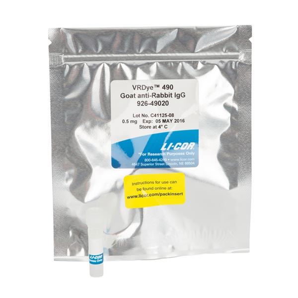
Highly cross-adsorbed goat anti-rabbit IgG (H+L) antibody conjugated to VRDye 490.
Immunogen
Rabbit IgGPurity and Specificity
Isolation of specific antibodies was accomplished by affinity chromatography using pooled rabbit IgG covalently linked to agarose. Based on ELISA and flow cytometry, this antibody reacts with the heavy and light chains of rabbit IgG, and with the light chains of rabbit IgM and IgA. This antibody was tested by dot blot and and/or solid-phase adsorbed for minimal cross-reactivity with human, mouse, rat, sheep, and chicken serum proteins, but may cross-react with immunoglobulins from other species. The conjugate has been specifically tested and qualified for immunofluorescence microscopy and flow cytometry applications.
Applications
- Microscopy
- Immunohistochemistry
- Flow Cytometry
Formulation
VRDye 490 secondary antibodies are supplied as purified immunoglobulin conjugates, lyophilized in phosphate-buffered saline, pH 7.4. Protect from light. Store at 4 °C prior to reconstitution.
Each vial contains 10 mg/mL BSA (free of IgG and protease) as a stabilizer and 0.01% sodium azide as a preservative, after reconstitution. Concentration is 1.0 mg/mL when reconstituted as directed. Refer to the pack insert for details on reconstitution.
Recommended Dilutions
| Application | Recommended | Suggested Range |
|---|---|---|
| Immunofluorescence Microscopy | 1:400 | 1:100 - 1:1,000 |
| Flow Cytometry | 1:1,000 | 1:200 - 1:2,000 |
| Other | User-optimized |
Suggested working dilutions are given as a guide only. Optimum dilutions will vary and should be determined empirically.
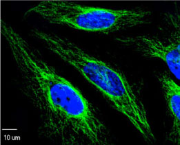
Example Data

